Categories

More Microscopic Images

In the lab, we have found more insects, a spider, and a lot of web in the Echinacea heads we have been cleaning. The insects include another lacewing fly and two beetles that I cannot identify. I also found a hard shell that looks like a Syrphid fly pupal case. I have the web and the egg sacs clustered because of the amount that we tend to find in the lab.

Unidentified Beetle No. 1:
Capture_00010.JPG
Capture_00011.JPG
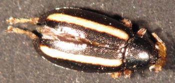
This beetle is black with two longitudinal yellow stripes down its dorsal side. It has wings and extended back legs for jumping. The mouth part is a golden color, similar to its legs. It also has reddish colored eyes. The beetle’s wings extend pass its abdomen slightly. The beetle also has very small antennae.

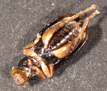
Unidentified Beetle No. 2:
Capture_00012.JPG
Capture_00013.JPG
This beetle’s body has been flattened in the bag or during cleaning. It is brown with lighter brown wings. The wings do not expand past its thorax. The beetle’s legs are short. The thorax and the abdomen form a pentagonal shape. The eyes are a dark brown color.

Lacewing Fly:
Capture_00018.JPG
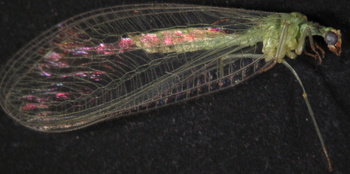
This lacewing fly has different characteristics in the abdomen than the previous one I posted. I suspect that the insects are different genders, based on the different abdomen shapes, but I was unable to find anything on sexing lacewing flies.

Spider:
Capture_00016.JPG
Capture_00015.JPG
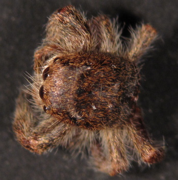
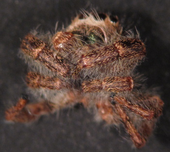
I suspect this spider to be a type of jumping spider due to the body and leg shapes and the fact that it has eight eyes. The four smaller eyes can be seen on the back of the head and this gives the spider more accurate vision and a wider range of sight. The spider’s head has a square shape with four eyes in the front and two eyes on each side. They are very hard to see, but there is a slight reflection of light where the other four eyes are located.

Syrphid Fly Pupa Case:
Capture_00017.JPG
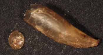
This is a hard, transparent shell of either a larva or a maggot that I could not identify. Stuart suspects that it is from a Syrphid fly pupa. The piece sitting next to it was broken off when I was moving the specimen with the forceps.

Spider Web Cluster:
Capture_00014.JPG
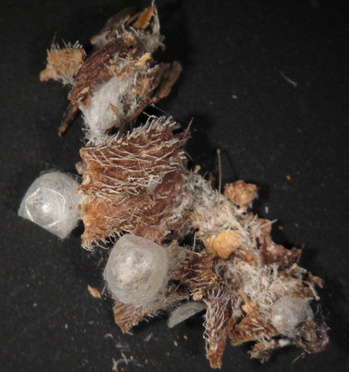
This is a cluster of web and egg sacs that we have found in the Echinacea plant material. We tend to find a lot of web and egg sacs when cleaning Echinacea heads.

planting Oenothera biennis seeds 14 Nov 2011

2:00 pm winds WSW

1 ew left/row [?]
Tape ends at 18.8m on the N side and 18.85 on the S side.
Plant at 0, 0.33, 0.67 m.
Flags at even meters (ending at 18.04).
Two Oenothera 1-yr olds at N edge 10.45m and 11.00 m.
Weeded Poa, Brome, Carduus, Cirsium, Euphorbia (before planting).
Zer[o] at westernmost grass.

[entered from notes in 2012; see plantOenotheraNov2011.pdf]

2012 Planned Common Garden Measuring Protocol

Here is the protocol that we plan to use for measuring in 2012:
2012.measureFieldProtocolPlan.htm

2011 Common Garden Measuring Protocol

Here’s a link to the protocol that we used for measuring CG1 in 2011:
2011.measureFieldProtocolReal.htm

In 2012 we plan to measure in “review mode” (as we did for CG2 in 2011) — all location records will be on the Visors with Status=”Staple” or “Skip” populated. We should not spend as much time searching for plants that have not been present for 3 or more years.This should speed up measuring. I’ll post the planned 2012 protocol next.

Taking Photos of Insects Using a Microscope

My name is Jill Meyer and I am a student intern working on the Echinacea Project. We were sitting and cleaning heads in the lab when I had found an exoskeleton of a molted grasshopper. It was a new and exciting discovery for me, as I did not previously know that grasshoppers molt. I have always been semi-fascinated with bugs, so I placed the grasshopper’s exoskeleton in the drawer to save. Since then, my collection has grown and I have added an Assassin Bug, a spider egg sac, a lacewing fly and a pentatomid. These can be found below. Unfortunately, the insects have fallen apart in the envelope I have kept them in and their limbs are captured laying next to their bodies. I have also written a lab procedure on how to take these images using a microscope, which I have also included below.
Pentatomid
Thumbnail image for Capture_00002.JPG
Spider Egg Sac
Thumbnail image for Capture_00004.JPG
Assassin Bug
Thumbnail image for Thumbnail image for Capture_00005.JPG
Lacewing Fly
Thumbnail image for Thumbnail image for Capture_00006.JPG
Grasshopper Exoskeleton
Thumbnail image for Thumbnail image for Capture_00007.JPG
Procedure:
Taking Images of Insects Using a Microscope Procedure.doc