|
|
We’re done! Yesterday Emma and I finished up our poster with mere minutes to spare and some timely help from Stuart (the man knows concise language). I thought of about five other tests that we should run (to characterize interactions between explanatory variables, etc) while wrapping up our last section, but defeated the urge. We’re both very proud of the poster and the work we did these past three weeks.
Thanks to everyone in the Echinacea Project for your guidance, help, and companionship! This externship has been an awesome experience and has definitely helped us to grow as scientists.
Here’s a link to our final poster:
Kropp_Stewart_Poster
After two grueling days and one exceptionally grueling one (during which we had to solve an issue with our tinies–what even is a confounding variable??), we’ve finally finished analyzing our data.
Our models fit, and our p-values are significant (sometimes), but the best takeaway from this week is definitely what we’ve learned about statistics and R. Prior to this Emma had never seen R before and I had only a rudimentary knowledge, but yesterday we wrote some code and solved a few problems without even consulting the internet! A HUGE shout-out to Stuart, Scott, and Amy for teaching us about generalized linear models, backwards step-wise regression, logistic transformations and all of the R syntax we didn’t know we needed, and for being patient with our sometimes-constant questions. I indubitably feel like I understand much more than I did coming into this week.
Stay tuned for our poster, which will include all the juicy juicy results that Emma and I have been slaving over. How has time passed this quickly?
Mikaela
Classifying x-rays stopped being fun very quickly today. Emma and I had divided the slides randomly and were each doing half, and I finished mine first so I decided to classify a handful of hers and see how the numbers matched up. They didn’t. Around 50% of the slides I recounted had new numbers. All of the discrepancies were because achenes were in between being full and partial or partial and empty (partial achenes were fertilized, but the embryo either did not develop normally or was destroyed). Together we went back to go over the discrepancies, and we came up with a few rules to make things clearer in the future. We were already working with a few rules that Danny had set out for defining partials:
- The embryo is fragmented
- The embryo has concave (curved inward) or irregular edges
- The width of the embryo is less than half the width of the achene at its widest pointThis was a good start, but we still found plenty of ambiguous achenes. Our new definitions for “partial” are:
- Both the top and bottom of the embryo is rounded, and there is plenty of space not filled within the achene. Looking for space along the sides of the achene can help identify this one.
 (71top)
- A strong, bright line inside an achene indicates a partial achene, whether it curves or not. Make sure this is distinct from the edges of the achene.
 (59bot)
- If the achene is bright, but not as bright as regular full achenes, the achene is partial (this can be described as cloudiness).
We also came up with a few hints (learned the hard way):
- Be careful of florets–they can show up as brightly as embryos, but don’t count them! The florets at top left of this image are almost as bright as the embryos bottom-right, and could be mistaken for partial achenes.
 (93top)
- If a bright spot has no outline of an achene around it and no band near it, and it isn’t a recognizable shape, assume it is chaff and let it alone.
- The “band” is the little bright line at the top of x-rayed achenes.
- When there is less contrast in an image and the brightness of embryos is hard to see, look at the bands at the tops of each achene. These are usually about as bright as embryos, so you can set a standard by that shade.
 (70bot) (70bot)
It is also worth noting that we decided to count achenes as full when the embryos don’t fill the entire outline: a space between the embryo and the achene is definitely permissible, and even smaller embryos count when they still maintain the proper shape (flat/convex top, pointed bottom) and aren’t smaller than half the width of the achene.
It was a long day of checking and rechecking classifications, but we’re finally happy with our counts. Now we’re working on a poster to help recruits learn the difference between informative and uninformative achenes, and Amy is helping us compile data. We’re officially out of the collection phase!
Always tired,
Mikaela
Whew! This week has been BUSY–and it’s only Wednesday! Here’s a run-down of our milestones:
Monday: FINISHED CLEANING SEED HEADS. For good. For ever. Cleaning is definitely the most time consuming step of our process, and having it over with is a dream. We also learned how to x-ray our seeds, and my feelings on it are pretty confused. On the one hand, radiation is scary; on the other, using x-rays for botany is pretty darn cool. On the third hand, there is a lot of waiting involved in the process. All in all, definitely glad I learned how to do it.
Tuesday: We finished scanning on Tuesday morning (all 110 scans!) so we’re feeling pretty good going into the rest of the week. In the afternoon we got a start on randomizing and met with a PhD student here, Jessa. She was really cool–we talked about grad school, biology and our names, which are all fun and interesting things to talk about (note that neither Emma nor I have gotten started on thinking about grad school yet–it’s only fun to talk about for now).
Wednesday: We finished randomizing! And then we finished assembling! And then we finished x-raying!! Then we did some data entry…and then we finished counting! Because we already processed 2/3 of our samples last week, this final pull has been a breeze. (Which Scott and Amy are blown away by. Apparently we’re pretty quick!)
My head is whirling a bit from everything we’ve accomplished this week, but we still have a lot to do. Our next steps will be:
- Finish classifying the x-rayed seeds (we got a start on this today and I love it; it could make a pretty good video game)
- Compile our data for analysis
- Analysis!
We’ve decided to focus our analysis on the uninformatives. Are smaller seed heads more likely to have tiny uninformatives? Are seed heads from dense populations more likely than isolated heads to have larval holes in their achenes? These and many questions are waiting to be answered…I’d better get back on that!
Mikaela
 These yellow coin envelopes contain the products of cleaning: achenes sorted into top, middle, and bottom for each seed head. The pink and blue cards at the front of each box show our progress. These white coin envelopes contain the products of randomization: the uninformative achenes from each yellow envelope. The informative achenes are in the clear baggies in the next image! Sheets prepared for x-ray. Each of the little baggies contains informative achenes from either the top, middle or bottom of a seed head.
This morning I was devastated to find that our larva had died overnight–as we said in the previous post, we were hoping to raise them, and I had also become a little attached. My grief didn’t last very long, though, because I found two larva wreaking havoc in my second head of the day! Emma and I quickly repopulated our petri dish with wriggly pink bugs.
When a seed head has a larva, we can expect a good number of the achenes to have holes burrowed in their sides. Today, we finally caught a larva in the act! This little guy was found with the front half of its body inside an achene. It’s good to have conclusive evidence for what precisely is going on around these seeds…
Other exciting updates: we’ve officially passed the half-way point on cleaning our 110 seed heads. At closing, we’re working on #61. Woo for progress!
Ta ta for now,
Mikaela
 The perpetrator (image quality fuzzy to better convey shadiness)
So it begins! Two new externs have joined Team Echinacea from Carleton College. We (Mikaela and Emma) will be here every day for the next three weeks, and are excited to discover new revelations for the Asynchrony, Isolation and Incompatibility experiment.
So far, most of what we’ve discovered is that cleaning Echinacea seed heads is tedious. Two days in, we have cleaned 36 seed heads; scanning them was a nice relief from the monotony. We think we could get through all 110 by the end of this week.
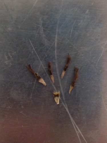
Although yesterday was quiet, there was a little bit of commotion: Mikaela’s second seed head had a rare deformity. Many of the achenes were uninformative. This means they were aborted part of the way through formation, so it cannot be determined whether they were fertilized. After minutes of puzzled deliberation, Stuart, Amy and Scott decided to keep them in the sample.
 Four uninformative achenes compared to one normal, small-to-mid-size achene. Because of their immaturity, the florets are still firmly attached. In contrast to yesterday, today there were quite a few volunteers and a couple of students who we got to meet. It was nice to talk to other people who were involved in and excited about this project. We also got to hear about other experiments going on in the lab besides our own.
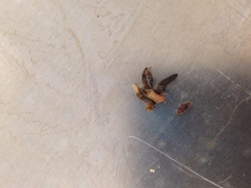
Today’s seed cleaning also presented an exciting moment: just moments after Amy told us about last year’s larval discoveries, we each found a live larva residing in the heads we were cleaning. We’re thinking about raising these mystery larva so we can finally learn just what they are. Hopefully we’ll have more success than last year’s effort!
Our two larva. Emma’s is the tiny brown one on the right, and Mikaela’s is the pink one hanging out on a makeshift habitat of chaff. We are grateful for this opportunity to contribute to and learn from the project, and are looking forward to the next three weeks!
Thanks for the warm welcome,
Mikaela and Emma
|
|


 (70bot)